3D IMAGING (CBCT)
Cone Beam CT (CBCT)
Cone Beam Computed Tomography (CBCT) is a relatively new technique, which is mainly used to assess the jaws and teeth. Its main advantage compared to OPGs and other dental x-rays is that it provides three-dimensional imaging (similar to conventional CT, but with a lower radiation dose). Nidaan uses an advanced Cone Beam CT (CBCT) system, which provides high definition, three dimensional imaging to complement our other dental imaging services like OPG. Cone Beam CT offers 3-D evaluation of dental anatomy and pathology: for example impacted teeth, pre-implant assessment, orthodontic assessment, and assessment for jaw and face surgery. The x-ray beam is cone-shaped and it generates high quality images. CBCT scan needs lower radiation, which is a huge advantage over conventional CT scan. High quality images from CBCT scan help dentists to determine the cause of the dental issue and plan for more precise treatment.
Procedure
The three-dimensional image data will also be provided on a disc to the dentist or specialist who referred you, and can be used with other software, for example for implant planning.
CBCT IN IMPLANTS
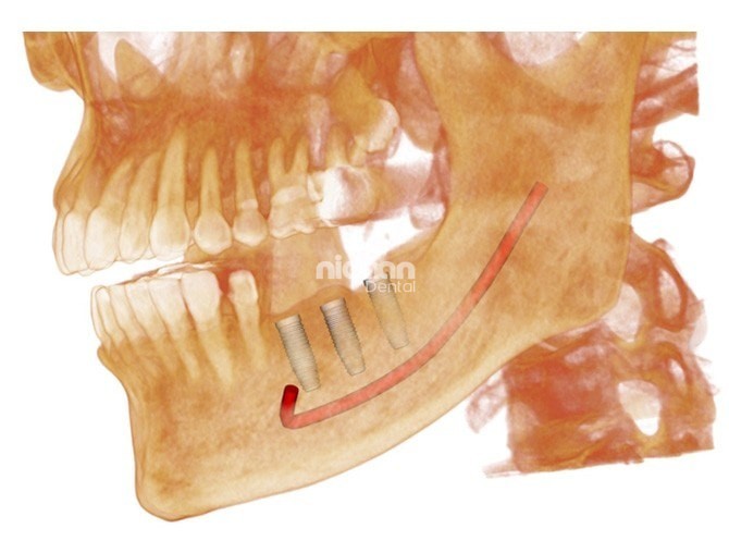
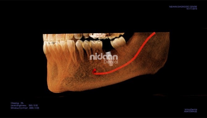
Study consists of cross sections and panoramic projections using Cone Beam CT (CBCT) scanner. It can be of an individual area or the whole mandible or maxilla. CBCT 3D technology is one of the most effective tools available for evaluating implant sites. 3D images can identify potential pathologies and structural abnormalities with unprecedented accuracy.
Cross sectional and panoramic views facilitate various measurements such as: height and width of the implant sites, mandibular edentulous site, a potential implant site near the mental foremen, width of the buccal/lingual ridge and cortical bone density. 3D images highlight the cortical bone thickness, the cancellous bone density, inferior alveolar nerve and the mental foremen location. They also influence the choice of the appropriate implant to be used, its placement, its width and consideration of “die back” from dense cortical bone.
CBCT IN TM JOINT EVALUATION
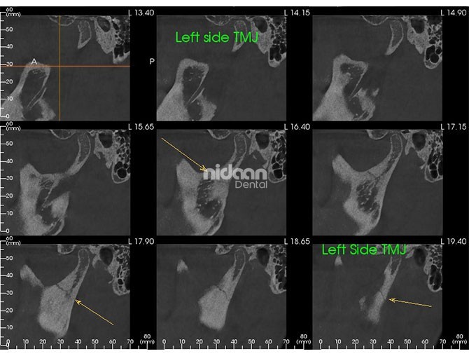
3D CBCT takes the examination of the Temporomandibular Joint (TMJ ) to a new level. After a single scan, Sagittal and Coronal views can be sectioned to show joint space and pathologies. 3D image reconstruction can clearly provide detailed information of the TMJ and Cervical Spine anatomy. A wide panoramic view provides a quick screening tool, where differences in condylar and ramus height as well as other dental pathologies can be checked.
CBCT IN ENDODONTICS & PERIODONTICS
In order to perform certain procedures, like treating a fractured tooth, mandibular canal therapy and caring for the surrounding tissue, endodontic and periodontic specialists require extremely high quality images that will allow them to identify every detail of the treatment area, make an accurate diagnosis, and establish an effective treatment plan. Upon carrying out a thorough examination of the area in question, the user will gain a full appreciation for the device’s less invasive nature and greater suitability.
CBCT IN ORAL AND MAXILLOFACIAL SURGERY
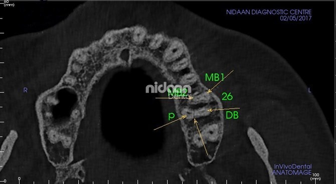
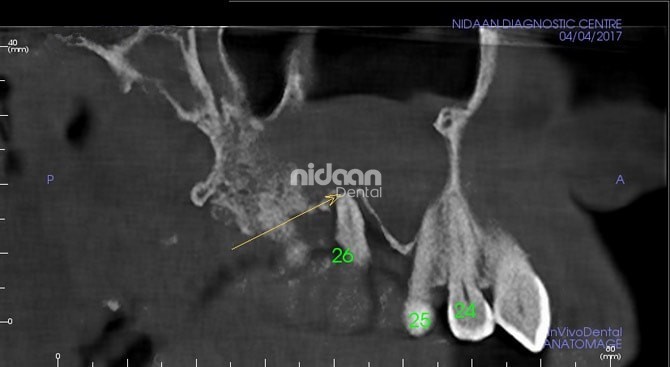
This discipline deals with the correction of various soft and hard tissue diseases afflicting the maxillofacial area. Scans performed precisely illustrate specific characteristics, such as the presence of teeth or fractures, bone density and depth, and the shape and the inclination of the root. Furthermore, in the case of post-operative scans, the presence of any metallic elements will not affect image quality. On the contrary, thanks to the low number of rays necessary, the scattering effect is almost non-existent, thus allowing the anatomical structures scanned to be clearly displayed.
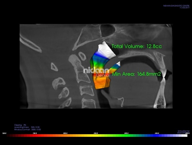
CBCT – Airway Assessment in Obstructive Sleep Apnea (OSA)
The most recognized risk factor for obstructive sleep apnea is an anatomical narrowing of the upper airway. In particular, the locus is in the oropharyngeal area. Identifying the exact location of the obstruction is important and upper airway imaging techniques can help with this question. Scan performed precicely illustrates the anatomical region of narrowing to guide the doctor in treatment.
